
About seventy years ago, a highly regarded neurosurgeon from Columbia, Dr. Solomon Hakim, noticed that on autopsy many patients with Alzheimer’s disease had enlargement of the ventricles without destruction of the outer cortex of the brain, which would have happened if the enlargement was due to high pressure. The ventricles are chambers in the center of the brain and brainstem where a watery substance called cerebrospinal fluid (CSF) is produced. The purpose of CSF is to support and protect the brain. It also removes waste from the brain. Enlargement of the ventricles is called ventriculomegaly. Ventriculomegaly seen on brain scans is a sign of hydrocephalus, which is an increase in CSF volume in the brain.
Ventriculomegaly stretches and deforms the surrounding periventricular structures. Prolonged deformation can lead to plastic deformation, which is permanent. Ventriculomegaly also compresses the veins, that are located on the outer surface of the brain, against the bones of the cranial vault. This can decrease venous drainage of the brain and cause the hydrocephalic condition to worsen. Ventriculomegaly and damage to periventricular structures may play a role in many of the signs and symptoms associated with neurodegenerative diseases such as motor weaknesses, dementia, cog fog, heat intolerance, sleep disturbances, sleep apnea and incontinence of the bowel and bladder.
 Dr. Hakim later called the condition he discovered normal pressure hydrocephalus (NPH). He also made a major improvement in the design of the spring on the valves that are used in shunts to treat hydrocephalus. The principle behind his modification to shunts is still in use today.
In 1976 Dr. Hakim published a paper in which he compared the brain to a sponge and suggested that poroelasticity plays a role in the development of ventriculomegaly. The term poroelasticity comes from engineering sciences related to soils and rocks and will be explained later in this post. Since the advent of brain scans, ventriculomegaly has been associated with Alzheimer’s disease, Parkinson’s disease, multiple sclerosis, and other neurodegenerative conditions. Mega cisterns are enlarged CSF chambers similar to ventriculomegaly and will also be discussed in this post.
In the brain scan below, the lateral ventricles are located beneath the large white arch-like structure. A smaller arch-like white band underneath the larger structure is joined to it in the rear and is the bottom of the lateral ventricle. The third ventricle is below the lower white band and above the midbrain which is the uppermost part of the brainstem (stalk-like). The fourth ventricle is the dart shaped area between the brainstem in front and a cauliflower-like looking structure in the rear, which is the cerebellum.
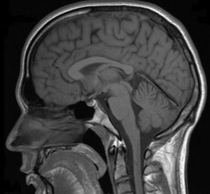 The dark gray spaces in front of the brainstem and beneath the cerebellum are called cisterns. CSF flows out of the fourth ventricle and into the cisterns. The cisterns cushion the brain from the hard walls of the cranial vault. They also support the brain and prevent it from sinking into the large hole in the bottom of the vault called the foramen magnum. This is the opening for the passage of the brainstem and spinal cord, as well as blood vessels and CSF pathways.
Ventriculomegaly can be caused by an increase in CSF volume and pressure in the ventricles, or it can be caused by atrophy (shrinking) of the brain due to degeneration of the structures that surround them. It can also be a combination of atrophy and changes in CSF volume and pressure. In many cases the cause of the ventriculomegaly is unknown. In any case, enlargement of the ventricles can affect the important structures that surround them in what is called the periventricular areas.
The periventricular structures that surround the ventricles are some of the most important and fundamental systems in the brain such as the limbic (reptilian/visceral/self-preservation) and autonomic (vegetative) nervous systems. The roof of the lateral ventricles is formed by a large group of myelinated (white matter) nerves that link the left and right hemispheres of the brain. Descending long myelinated motor (muscle) nerve tracts called the internal capsule pass close to the ventricles.
To get a better understanding of the mechanical forces that can cause enlargement of the ventricles, researchers have been turning to engineers, mathematicians and physicists for answers. From an engineering perspective, the skull and brain, as well as the fluids inside them can be compared to rocks and soils. Poroelasticity is a property of rocks and soils that affect their structural strength and their abiltiy to support large loads such as from water, waves, buildings, bridges and roads. Different types of rocks and soils, as well as the shapes of their pores, fissures, fractures and caves affect the way they handle loads. Similarly, poroelasticity affects the structural strength and shape of the brain, as well as deformation such as ventriculomegaly. This is important because deformation of the brain such as enlarged ventricles can damage nearby delicate nerves and blood vessels. Damage to nerves and blood vessels can, in turn, lead to atrophy (shrinkage) and ventriculomegaly.
 The term poroelasticity refers to the pores in soils and rocks that affect their elasticity. Elasticity is the ability of a structure to deform and return to its original shape without breaking (fracturing). The pores in soils and rocks can be filled with gas or fluids. The gas could be air or natural gas. The fluid could be water or oil. The gases or fluids affect the strength of soils and rocks, as well as their elasticity. Consequently, the gases and fluids affect the ability of soils and rocks to deform and reform. Structures that go through expansion and contraction are considered to be biphasic. If it can’t deform and reform, meaning return to its original shape, the structure is considered to be monophasic. The ability of a structure to deform and return to its original shape is determined by, what is called, its elastic coefficient.
As far as biphasic poroelastic properties are concerned, blood and CSF are essentially non-porous and non-compressible. They also lack elastic properties. Instead, they have viscoelastic properties that are entirely different. Basically, elasticity is a property of solid structures. Viscoelasticity refers to properties of liquids and foams such as viscoelastic memory foam mattresses used for sleep surfaces. In contrast to blood and CSF, the brain is made of billions of cells that are filled with fluids called intracellular fluids. The brain’s many fissures, sulci, interstitial spaces, perivascular spaces, subarachnoid spaces, ventricles, caverns, cisterns and sinuses are all filled with fluids such as, intracellular fluids, interstitial fluids, blood and CSF. This makes the brain a highly porous liquid filled structure.
In addition to being porous, the brain is also elastic. For example, tumors and hydrocephalus can cause significant deformation of the brain. When the stress is removed, however, it returns to normal size provided permanent damage has not yet occurred. Being elastic technically makes the brain a biphasic structure capable of expansion and contraction. Under normal circumstances, however, the brain is only slightly biphasic. This is because it is completely surrounded and all it spaces are filled with CSF. CSF pressure causes internal and external tension, called turgor, in the pores and spaces of the brain. Turgor causes stiffness.
The stiffness caused by turgor is important to maintaining the shapes of living things. Plants use turgor to stay upright. If they become dehydrated they quickly start to droop. Cells similarly use turgor to maintain their structure and internal space and prevent compression. Turgor, likewise, maintains the shape of the brain. Maintaining the shape of the brain is important because it prevents compression of delicate nerve structures and smaller blood vessels that travel through sulci (folds), fissures (cracks) and foramen (holes) in the brain and skull. Turgor also keeps the ventricles from collapsing which is called slit ventricles. Slit ventricles occur due to overdrainge of CSF by external surgical shunts. They can also occur when the normal CSF pressure gradient is reversed. I will discuss shunts, siphons and slit ventricles further in future posts. In addition to maintaining the size of the ventricles turgor helps to keep the brain afloat and prevent it from sinking or making contact with the cranial vault.
Turgor has its limitations, however. Too much turgor limits the compliance of the brain. Compliance is a term used to describe the stretch phase of elasticity in the brain. Compliance allows tissue to deform without damage such as with compression and stretching. Elastance is the complete opposite of compliance. Elastance is the strength of tissues to resist deformation and return to their original shape once the load is removed. Elastance preserves the designs and shapes of structures. On the other hand, the compliance of certain tissues in the brain, especially weak-walled veins, allows them to buffer the impact of the relatively large volume of blood and the associated increase in pressure that is pumped into the cranial vault with each contraction phase of the heart roughly seventy times per minute. The strength of the arterial waves of blood and the pressure pumped in during each heart beat needs to be decreased and modified before sending it into the delicate internal structures of the brain.
The dark grey areas on the bran scan above is CSF. As you can see, CSF fills all the spaces, cracks and caves. The brain is also contained inside the cranial vault and surrounded by CSF that maintains its position and prevents contact with the bones of the vault. Although it is highly porous, the interior and exterior surfaces of the skull are more like limestone or granite in that it has poor permeability. The non-permeable hard shell of the skull protects the brain and keeps the weather out. Although it lacks permeability, the skull is penetrated by many holes called foramen and other openings, which makes it highly porous. Nerves, blood and CSF travel through these openings. Moreover, pressure in these openings can have a profound effect on fluid mechanics in the brain.
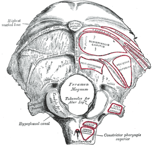 While it is not considered to be a factor that directly affects compliance (elasticity) in the brain, the foramen magnum and spinal canal play an important role in maintaining CSF volume and pressure in the brain. As shown in the picture on the left of the skull base, the foramen magum is the large hole in the base of the skull that connects the cranial vault to the spinal canal. The arterial pulsations and pressure waves that are pumped into the brain by the heart are not only buffered but a proportionate outflow in the amount of blood and CSF is also transferred out of the cranial vault and brain through the foramen magnum and into the spinal canal.
Obstruction of venous and CSF pathways in the foramen magnum can affect intracranial pulsatility and pressure waves. In particular it can cause back pressure on the drainage system that results in increased turgor resulting in stiffness due to loss of compliance (elasticity). Obstruction to blood and CSF flow through the foramen magnum can also cause CSF inversion (reverse) flows, turblulence and water hammers in the brain. CSF inversion flows, turbulence and water hammers may play a destructive role that results in damage to periventricular structures.
The most common place for obstruction of CSF outflow to occur is in the craniocervical junction (upper cervical spine). The most common causes of obstruction in the craniocervical junction are malformations and mechanical strains such as misalignments. Obstructions due to malformations and mechanical strains indirectly but significantly affect intracranial compliance (elasticity) and the ability of the brain to absorb and control fluid mechanics caused by heart contractions.
To get a better understanding of how faulty fluid mechanics batter the brain, researchers and engineers are now plugging biphasic poroelastic properties of the brain into computational fluid dynamics and finite element analysis formulas to form computer models to predict and determine the cause of ventriculomegaly. They are also using physics formulas for computational fluid dynamics to determine flow through the different structures of the brain such as the ventricles. The different structures of the brain and the skull have complex shapes and different materials with different degrees of strength and compliance (elasticity), as well as their differing response to hydraulic pressure.
Hydraulic force is a product of pressure multiplied by the size of the area the pressure is being applied to, such as the volume of the ventricles or cisterns for example. A hydraulic pump can be used to increase force by applying pressure to a larger cylinder. Similarly, the pressure from the heart exerts more force on the larger pores and spaces of the brain compared to smaller ones. In this regard, the largest spaces in the brain are the ventricles and cisterns. The effects of hydraulic force may play a role in the ventriculomegaly seen in neurodegenerative condtions such as Alzheimer’s, Parkinson’s and multiple sclerosis. Constant strain from increased force in the ventricles and cisterns may cause a breakdown in the elastic properties of the brain so that the ventricles and surrounding structures can no longer return to their original shape, which is called plastic (permanent) deformation. Dr. Hakim also suggested, many years ago, that the greater size of the ventricles allow them to exert more force and thus maintain the ventriculomegaly with relatively lower pressure (turgor).
In addition to Hakim’s theory regarding ventriculomegaly, my theory regarding mega cisterns, which are enlarged cisterns, is that they are caused by inversion flows, turbulance and hydraulic forces that damage the brainstem and cerebellum resulting in atrophy of nearby structures. Mega Cisterns and atrophy of the brainstem are seen in mega cisterna magna, the Dandy-Walker Complex-Continuum, and a variant form of Parkinson’s disease called olivopontocerebellar atrophy, also known and Shy-Drager Syndrome or Multisystem Atrophy. Understanding these conditions will shed further light on the role of faulty fluid mechanics and hydraulics in neurodegenerative conditions of the brain. I have covered these conditions previously and will cover them more in future posts.
Hydrofracking is a term method engineer’s use to fracture rocks with water pressure. A similar situation called a water hammer can occur in the brain and damage delicate tissues. The location of the periventricular tissues predisposes them to compression, shear forces and water hammers that can cause damage. Loss of compliance in the brain magnifies the destructive forces. My next post will be on hydrofracking and brain atrophy (shrinkage).
For further information on enlargement of the ventricles and autonomic dysfunction called dysautonomia visit my website www.upright-health.com.

Thanks Dr F,
after reading this I see an importance of regular eg annual MRI’s to keep a record of the Brain mass so that issues such as Atrophy are monitored.
The dx process for the conditions you are describing would need very accurate observations of the brain regions to achieve the precise dx.
I have noticed over the years as MRI techniques change and the dx’s are made through different techniques with the MRI’s that monitoring for example the Ventricle sizes would be difficult, each set of MRI slides I have are so different that one would be hard pressed to say they are of the same person.
Another great article and more brewing!
Nigel
Your welcome Nigel,
Thanks for the compliment. You are becoming whiz on the subject.
Although it would be nice from a scientific point of view, I don’t think we need to monitor ventricle and cistern sizes in every patient or frequently. The cases that require closer inspection and monitoring are those with signs of suggestive of enlarged ventricles and cisterns. Some cases of increased intracranial pressure have normal sized ventricles and cisterns due to increased resistance (turgor) in the subarachnoid space that prevents them from enlarging. Increased intracranial pressure can also be associated wit slit ventricles if the CSF pressure gradient is reversed and pressure in the subarachnoid space is higher than the pressure in the ventricles.
Hi there, is MRI the most accurate and cost effective method for measuring Ventriculomegaly? Is there anyway we can test for it in practice apart from cranial nerve testing etc
Adrian
Hello Dr. Clegg,
Brain scans are the only way to check for ventriculomegaly. The absence of ventriculomegaly, however, doesn’t rule out hydrocephalic conditions. For example, an increase in transmantle pressure due to sluggish CSF flow, in which CSF accumulate outside the ventricles prevents ventriculomegaly. It is better to monitor the patients signs and symptoms, including the cranial nerves.