
Researchers suspect that enlarged ventricles, known as ventriculomegaly, seen in many neurodegenerative diseases may be the result of atrophy (decrease in size) of the brain. The cause of the damage or atrophy of the brain may be due to destructive waves and hydraulic pressures that damage tissues by a process I compare to hydrofracking which is used by engineers to fracture rocks. Ventriculomegaly and brain atrophy have been associated with Alzheimer’s disease, Parkinson’s disease and its variants, called Parkinson’s Plus, as well as multiple sclerosis, amyotrophic lateral sclerosis and Huntington’s disease.
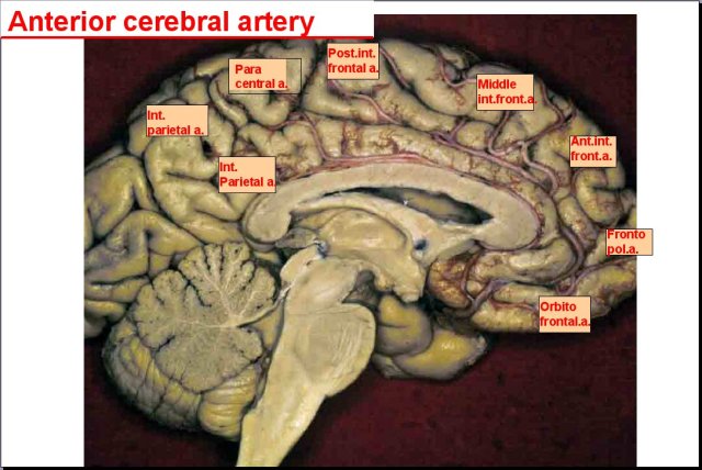
The picture above shows the left half of the brain. The face would be to the right. The cauliflower structure in the lower left corner is the cerebellum. The hollow area (darker grey) in the middle of the brain is the left lateral ventricle. The heavy white structure that forms the roof over the lateral ventricle is the corpus callosum. The heavy white structure that forms the floor is the fornix. The corpus callosum is a group of myelinated (white matter) high speed interconnecting communication pathways that link the left and right halves of the brain. The fornix is, likewise, a high-speed communication pathway of white fibers. The third ventricle is located just below the fornix. The fourth ventricle is the space shaped like a dart between the cerebellum and brainstem. The ventricles are chambers in the core of the brain and brainstem where cerebrospinal fluid (CSF) is produced.
CFS is basically water with some sugar and a few other ingredients mixed in. CSF fills the ventricles and surrounds the entire brain in a water jacket. CSF in the ventricles, fissures and spaces of the brain serves to cushion and protect the brain from compression against the bones of the cranial vault, as well as maintain its shape, layout and position inside the vault. It also serves as the lymphatic waste removal system of the brain. Due to the constant state of tension caused by CSF in the ventricles and spaces in and around the brain, some engineers consider the brain to be essentially a non-compressible monophasic structure. Monophasic simply means that it doesn’t buckle and deform under pressure.
In contrast to engineers, chiropractic and osteopathic craniosacral theories have maintained for many years that the musculoskeletal system, CSF and central nervous system, which includes the brain and spinal cord, rhythmically pulsate and move. The movement and pulsations are driven by neurological, cardiovascular and respiratory waves. More recently, radiologists have similarly shown that CSF pulsates and that the ventricles expand and contract in synchrony with cardiovascular waves. They have also shown that the brain moves up and down like a piston during each cardiac cycle. This is because the increase in volume, mass and pressure of the brain caused by the increase in blood volume drives the brain downward in the cranial vault. Relaxation of the heart relieves the pressure and strain, which causes the brain to rise inside the vault. This expansion and contraction coupled with up and down movement of the brain inside the vault makes it a biphasic structure in engineering terms and futher confirms the craniosacral theory.
The picture below is from an article published in 2011 in the Delaware Free Media News on the politically controversial process called hydrofracking. Hydrofracking is done by pumping water under pressure into rocks located deep below the surface of the earth to open their fissures (cracks) and pores by fracturing them. The process is mostly used to flush out oils and gases to be brought to the surface and refined. In addition to the potential pollution of water aquifers that are located above the fracture zone, the compression and shear stress caused by hydrofracking can set off vibrational waves deep below in the fracture zone that travel to the surface where they can be felt as earthquakes by residents. The earthquakes can cause tears in the surface of the earth and cracks in buildings.

A similar situation can occur inside the cranial vault of the skull due to the heart pumping a relatively large volume of blood with each contraction, into the mostly closed container of the cranial vault. If the blood volume and pressure coming into the cranial vault isn’t sufficiently buffered (within the subarachnoid space shown below) before it enters the brain, strong pressure waves can be sent into the core of the brain. These high pressure waves can damage delicate tissues resulting in atrophy and subsequent ventriculomegaly. The structures that most often show atrophy are often located in the periventricular areas, the areas that surround the ventricles.
Aside from atrophy, some cases of ventriculomegaly are caused by an increase in CSF volume due to obstruction or faulty flow. In these cases, the ventricles and brain return to normal size when flow is restored via shunts and surgery due to their biphasic nature. While these cases are seen much less frequently thus far, early detection may change things and prevent permanent damage and subsequent atrophy.
In addition to unchecked incoming high pressure arterial waves causing problems, damage can also occur to periventricular structures due to overstretching similar to an overinflated balloon. At the same time, the expanding ventricles can compress neighboring structures and blood vessels. Compression of blood vessels can decrease blood flow in smaller blood vessels resulting in chronic ischemia and subsequent atrophy. Some researchers further suspect that faulty CSF flow may cause water hammers(explained below) in the brain similar to tremors and earthquakes caused by hydrofracking. Water hammers can similarly fracture surrounding tissues resulting in atrophy. In any case, the cause of the ventriculomegaly and atrophy can come from water pressure problems inside the ventricles or outside of them.
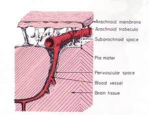 In contrast to hydrofracking which pumps large volumes of water into deep subterranean rocks, the heart pumps a relatively large volume of blood into the arteries contained within the subarachnoid space (see picture to the right). The subarachnoid space surrounds the outer surfaces of the lobes, the convolutions (gyri) and fissures of the brain and the brainstem within the cranial vault. The large incoming arteries pass through the subarachnoid space to supply numerous smaller branches (arterioles) that exit the subarachnoid space and enter tunnels called perivascular or Verchow-Robin spaces to supply smaller branches that supply the parenchyma (substance) of the brain.
As stated above, the periventricular areas are those structures that interface with the ventricles. These are important nerve centers. Blood vessels also pass through the periventricular space between the ventricles and surrounding structures. As you can see in the picture of the brain at the top of the page, arteries pass over the lateral ventricles. Veins also pass over the lateral ventricles. Smaller arteries and veins are similarly located next to the third and fourth ventricle. In contrast to the veins on the surface of the brain, the periventricular veins are much smaller and more susceptible to compression. Smaller arterioles can, likewise, be compressed.
The stress from the increase in blood volume causes mechanical strains and temporary deformation of the brain as the subarachnoid space balloons slightly outward. In cases of high intracranial pressure, the areas of the subarachnoid space located near the bones of the cranial vault can compress surface veins of the brain against the bones of the vault and, thereby, decrease blood flow. Ballooning of the subarachnoid space also causes compression loads on the lobes of the brain and the ventricles similar to squeezing a sponge. Due to their location in the core of the brain, the periventricular areas are the most vulnerable to compression and shear stresses.
In addition to compression loads caused by enlargement of the subarachnoid space and ventricles, ventriculomegaly also causes shear stresses due to stretching of the periventricular structures and blood vessels. The combination of excess compression and shear stresses can, over time, cause mechanical damage to the structures and blood vessels, as well as decrease blood flow that can result in tissue atrophy (shrinkage).
The rhythmical beating of the heart thus causes pulsations and pressure waves to form in the brain, blood and CSF. Those pulsating hydraulic waves dissipate through the entire brain. If the pressure isn’t reduced appropriately, the high pressure arterial waves on the surface of the brain get directed inward toward the weaker more vulnerable parts of the brain, such as those surrounding the ventricles in the periventricular areas. These structures get compressed against the unyielding walls of the cranial vault on the outside and the stiff walls of the ventricles in the center of the brain that are supported by internal tension from CSF pressure. Chronic pulsatile high pressure waves can lead to hydraulic fracturing of vulnerable tissues. This can result in degeneration and atrophy of surrounding periventricular structures. Some researchers suspect that myelinated nerves (white matter) such as those that surround the ventricles, are more vulnerable to tension strains and subsequently more likely to tear (fracture) from excess loads.
In a healthy brain, the subarachnoid space typically buffers the increase in blood volume and pressure. Most of the force is absorbed by veins in the subarachnoid space which have weak walls and are easily compressible. Compression of the veins moves blood out of the brain reducing pressureby removing volume inside the cranial vault. Pressure is further relieved by squeezing a proportionate amount of venous blood and CSF out of the brain and cranial vault by way of the foramen magnum, which is the large hole in the base of the skull for the passage of the brainstem and cord. As the arterioles relax following contraction of the heart, and the arteries and veins begin to return to their previous size, the fresh supply of arterial blood in the subarachnoid space is released into the perivascular spaces of the brain under lower pressure.
 Blockage of venous blood and cerebrospinal fluid anywhere along their pathways can alter CSF flow and cause abnormal pressure waves. The abnormal pressure waves are the result of incoming arterial blood flow and pressure waves running into resistance from venous backpressure and reduced or blocked CSF outflow causing what plumbers refer to as a water hammer. As shown in the sketch on the left, water hammers occur in domestic plumbing when water flow out of a faucet is suddenly shut off. This causes waves to be reflected backwards and crash with incoming waves. Since stiff non-elastic pipes can’t absorb the force like elastic veins, it causes them to shudder like a tremor. The tremor causes the pipes to bounce on surrounding structures resulting in noises that sound like someone hammering on the pipes.
Alzheimer’s and other neurodegenerative diseases seen in adults are often associated with normal pressure hydrocephalus (NPH) in which the ventricles enlarge but CSF pressure remains normal or just slightly elevated. To this day, it remains a mystery to scientists as to how the ventricles can enlarge when CSF pressure is normal. The only plausible explanation so far is atrophy. In other words, the brain decreases in size around the ventricles creating space allowing the ventricles to enlarge when CSF moves into the ventricles.
Some researchers suspect that brain atrophy is caused by water hammers that damage susceptible tissues. Others suggest that it is due to compression and shear stresses mentioned previously. Still others suggest that compression and shear forces can damage blood vessels and decrease flow resulting in atrophy. Lastly, some cases are due to an increase in CSF volume due to faulty flow without atrophy. In other words, the brain is simply compressed and returns to normal size when CSF flow and volume are restored as mentioned above.
In brief, the biphasic brain is trapped between a rock and a hard place. The rock is the skull that surrounds and protects it. The hard places are the ventricles located in its core, as well as the surrounding spaces, filled with CSF. Shear stresses caused by stretching from ventriculomegaly strain the periventricular tissues. Increases in arterial blood volume during heart contractions cause compression load strains that deform the brain and ventricles. Abnormally high blood and CSF pressure waves coupled with water hammers compound the internal and external ventricular stresses and strains with tremors. Over time, strong chronic tremors can tear tissues.
Researchers are now looking into the impact of blockage of the venous drainage system of the brain and abnormal CSF pulse waves. Over time, chronic venous drainage problems and high CSF pressure waves can lead to hydrofracking, ventriculomegaly and atrophy of the brain. One of the most likely points of blockage to venous blood and CSF flow is in the cervical spine, especially the upper cervical spine. The most vulnerable structures to hydrofracking and subsequent atrophy are located in the periventricular areas that interface with the lobes of the brain.
For additional information on these and related topics visit my website at www.upright-health.com.

How important will the health of the arteries and veins be?
It seems that people are suggesting that endothelial layer is very important in the health of walls and that diet, exercise and mindfulness are crucial as a first issue in this hydraulicing system.
If there was infection and inflammation happening as so often is today then that and cholesterol would be an indicator of Brain vascularture health?
Regards,
Nigel
The health of the areteries and veins are important to the health of the brain. The endothelial layer is critical to the integrity and function of the blood vessels. Degeneration and subsquent stiffness of blood vessels can lead to hypertension and increased pressure waves in the brain. It can also cause strokes. Vascular degeneration is a cause of Alzheimer’s.
Signs of infection, inflammatory markers and cholesterol are helpful but indirect assesments of vascular health. Patients with healthy circulation can get infections. Patients with poor circulation and stasis are predisposed to infections. Better signs of vascular health are blood pressure and pulse testing along with thorough physical examination for tongue, eye, and skin color, as well as signs of edema and checking pulses in the extremities. Vascular imaging and flow studies are likewise more direct and better than blood chemistry tests.
Off topic, but didn’t know where to write….
Need some direction. Was diagnosed in 1990 with PPMS…never believed I had MS. MRI’s every second year were peppered with contradictions. Diagnosed with TMJ in 1988 as a result of trauma to head and neck…C1 through C6 thermal scan revealed asymmetry, not really problematic until 1997. Now at age 63, had CCSVI twice with minimal improvement in the form of pain relief. Have severed ties with neurologists and MS clinic since 2008. Want to pursue chiropractic and TMJ treatment. Am I too old? By the way, I have been confined to a wheelchair for 14 years. Pain medications for neuropathic pain have never worked. Do you have any suggestions about where to start? Hoping to hear from you.
Hello Patricia,
There may be a connection between the trauma to the head and neck in 1988 since your diagnosis of questionable MS was about two years later in 1990. You are never too old for quality chiropractic correction and care of the spine. The problem is the length of time you have had the problem and the many years you have been in a wheelchair. There is most likely some permanent damage to the spine and nervous system. Prolonged wheelchair use also causes further degenerative changes in the spine. Nonetheless, it is well worth pursuing care for your TMJ and spine to see if you can do something about relieving the pain maintaining the spine as much as possible. I don’t know enough about your case to make specific recommendations about where to start. I do offer a complimentary cursory consultation with the purchase of my book through my website that includes my email address if you are interested in providing more personal information about your case and where you live so I can see if I can help you.
Recent MRI report states, “There is evidence of abnormal diffusion/restricted water flow at several locations; specifically, right frontoparietal margin, right corona radiata, right fornix at the parietal occipital margin, and left forniceal region.” I was Dx with MS 21 yrs ago. has been a problem for last 6 yrs. Neurologist does not care abt the excess fluid in my brain. She didnt mention it during my post MRI visit. After reading your articles I’m wondering if this is normal hydrocephalis? Will you please steer me in the right direction? Thank you.
Hello Nancy,
From what you have told me, you have restricted water flow (diffusion) so you most likely have edema not normal pressure hydrocephalus per se. NPH, however, is essentially edema of the brain. You need to work on improving arterial blood flow to the brain and removing restrictions to venous and CSF outflow.