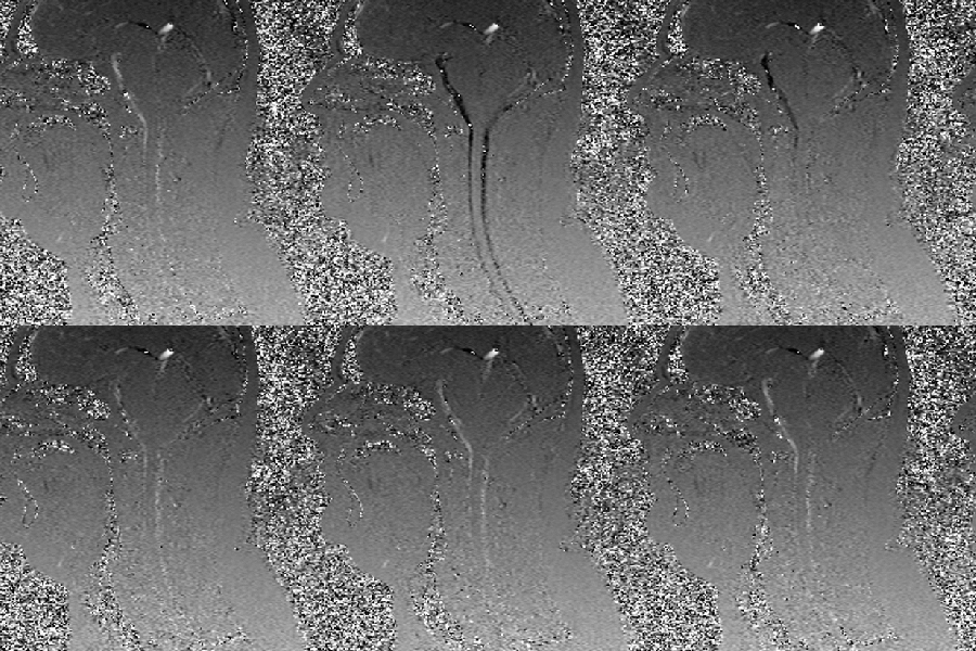
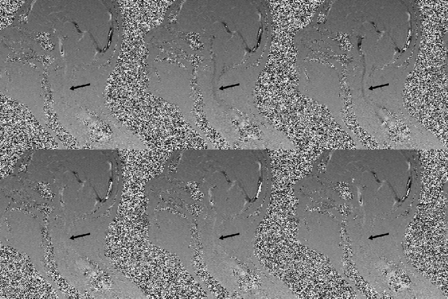
 The unusual images on the left and below were taken of the head and neck area looking at the patient from the side. The face is on the left side of the image. The images are called cine MRI because they are similar to cinema photography. In cinema photography many still pictures are taken and played back at high speed, which make the images appear to be moving.
In this image the speckled black and white areas are air surrounding the patient, as well as air in the paranasal (air) sinuses, the nose, the mouth and the throat. The more uniform gray area is the patient’s skin, fat, bones and muscles. If you click on the image you will see the movement of cerebrospinal fluid (CSF). The streaks of black that quickly appear and disappear in the moving image is cerebrospinal fluid (CSF), which is mostly water with sugar and some other elements mixed in. Although this particular image shows CSF flow, cine MRI can also be used to show blood flow.
These cine MRI images are from the FONAR Corporation. FONAR Corporation was founded by Dr. Raymond Damadian who was instrumental in the development of today’s modern MRI scanners. More recently, Dr. Damadian invented the upright MRI, which is going to change the way we see injuries, degenerative changes and diseases of the spine, brain and cord. The images are from a study by Dr. Damadian of eight patients with multiple sclerosis called, The Possible Role of Cranio-Cervical Trauma and Abnormal CSF Hydrodynamics in the Genesis of Multiple Sclerosis, published in the journal of Physiological Chemistry and Physics and Medical NMR in 2011. All of the eight cases were associated with trauma and all of them showed obstruction of CSF flow. The image below on the right is one of the cases.
The problem with conventional MRIs is that they are taken with the patient lying down. The problem is that upright posture causes significant changes in the mechanical loads acting on the spine, as well as fluid flow in the brain and cord. In contrast to humans, in most mammals the primary forces acting on the spine, while standing on four legs, are tension and shear stresses. In humans, upright posture primarily causes compression loads on the segments of the spine. Compression causes the segments of the spine to cave, bulge and slip in different directions when there is instability. Many conditions of the spine, brain and cord appear normal or acceptable when an MRI is taken with the patient lying down because the abnormalities only show when the patient is upright. Dynamic or kinematic upright MRI takes images in different positions, such as foward or backward bending of the spine. Dynamic MRI images picks up even more problems that might otherwise be missed by upright MRI alone.
As mentioned above, cine MRI images use fast moving still pictures that are taken in pixels and digitized for computerized imaging. The cameras are gated. Gated simply means that the MRI cameras are timed to take the images at particular intervals. In this case they are gated (timed) to take images based on the beat of the heart. In these images, CSF (on it’s way out of the brain) appears as flashing streaks of black on the front and back side of the brain and cord. It then quickly disappears. The black streaks show up when a large volume of blood is pushed into the cranial vault and brain by the contraction of the heart. As it does, an equal amount of venous blood and CSF is driven out of the cranial vault and into the spinal canal and cord. If you study the images closely you will notice that the image at the top of the page shows a clear continuous and uninterrupted normal pattern of CSF flow between the brain and cord, as well as a champagne glass shape to the passages. Click on the image below and compare the differences.
In the moving image you see black streaks the same as in the normal cine CSF flow study above, but the streaks are interrupted in certain areas or not as dark. If you look at the area the black arrow is pointing to you will see that the dark streak suddenly stops compared to the normal cine CSF MRI image above. If you look for the shape of the champagne glass you will see it is barely noticeable or missing on the back side. Instead, there is a dark streak with intense white streaks within located at the top of the champagne glass in the back of the brain. Those mixed black and white streaks are out of synchrony with the normal flow of CSF and they are probably going in the wrong direction. In other words, they are turbulant inversion flows or backjets of CSF in the brain. They could also be standing waves, or as kayakers prefer to call them, clapotis.
The dural sinuses (veins) that drain blood from the brain during upright posture follow a similar course to the CSF seen as black streaks in these images and empty into the vertebral veins inside the spinal canal. When the heart contracts, venous blood is driven out of the cranial vault along with CSF and both must exit together through the foramen magnum in the base of the skull and into the spinal canal. The vertebral veins are located between the walls of the spinal canal and the outer covering of the cord called the dura mater. The space is thus called the epidural space. CSF passes through the subarachnoid space inside the cord. Therefore, if venous blood flow becomes obstructed in the epidural space at or below the foramen magnum in the cervical spinal canal, the hydraulic pressure that follows is transmitted to the subarachnoid space of the cord and can affect CSF flow.
It has been my theory for the past 30 years that the blockage of CSF flow is most likely one of the root causes of neurodegenerative diseases of the brain such as Alzheimer’s, Parkinson’s, and multiple sclerosis. A decrease in CSF flow can affect the brain in several ways. One of the ways I suspect it causes damage to the brain is from backjets (and turbulance) of CSF in the cisterns of the brain. The cisterns are on the front and back side of the brain stem and cerebellum. The outline of the cisterns in these images is the champagne glass shape. Over time, chronic CSF backjets, turbulance and standing waves can compress and damage the brain.
There are many conditions of the spine that can affect blood and CSF flow in the brain and cord. For further information on increased CSF volume and backjets in the cisterns check out my website page on atrophy dysautonomia. Or visit the website at www.upright-health.com.

Who does these Dynamic Kinenetic MRIs , a Chiropractor or Neurologist?
Hello Nancy,
Either one them can order the tests.
We live in Montana. Do you know where I can get an CINE MRI done? Tim
Hello Tim, Your neurologist should know where the nearest facility is. If not, I suggest you call the FONAR Corporation for locations.
I live in Raleigh, NC. I have CSF fluid running out my nose, along with severe thoracic and cervical issue. When I stand or sit up straight, my mid back area starts killing me. Then my left ear, and sometimes both ears do their Darth Vadar thing, headache, weakness in arms and legs, and CSF fluid runs a lot worse from my nose. Who do you recommend I go see?
Hello Brenda,
What type of severe cervical and thoracic issues do you have? There are many types of cervical and thoracic issues that can cause ear symptoms, headaches and weakness in the arms and legs. Certain types can also affect CSF flow between the cranial vault and spinal canal causing CSF volume to increase in the cranial vault.
There are certain types of gentle, non-force chiropractic methods that are good for severe cervical and thoracic issues, such and the Cox Method of flexion-distraction with a movable headpiece. It all depends on what your particular issues are. The link below is to a youtube presentation of cervcial flexion-distraction. As you can see, it is very gentle. The table is very versatile and effective in treating many types of severe issues in the spine from head to tail.
Cox Cervical Distraction
The link below is to Dr. Schievink’s website. Dr. Schievnik is an expert on CSF leaks who might be able to point you in the right direction regarding the leaks. Again, I think it’s important however, that you investigate a possible connection between the cervical and thoracic issues and faulty CSF flow between the cranial vault and spinal canal.
Spinal CSF Leaks